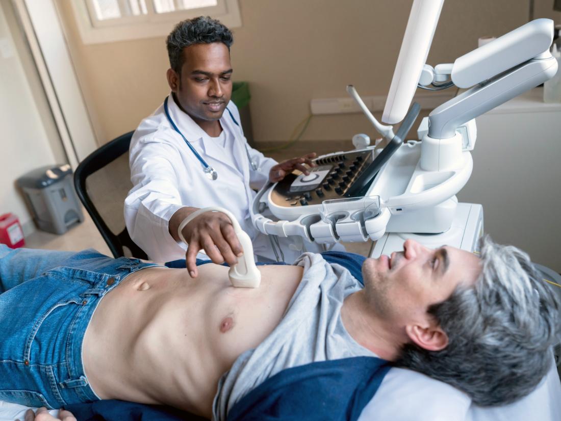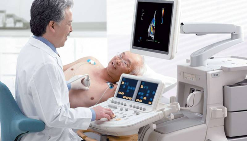
To identify heart diseases, your health care provider will use an electrocardiogram test to evaluate the structure and functioning of your heart. Board-certified cardiologist Sanul Corrielus, MD, MBA, FACC, and his team at Corrielus Cardiology perform echocardiogram in Philadelphia to evaluate the electrical activity in your heart, enabling them to detect diseases and determine the ideal treatment for you.
Understanding what an Echocardiogram is
An echocardiogram is a diagnostic tool that uses sound waves to produce images of your heart. Health care providers use the images from an echocardiogram:
- To analyze the beating of your heart
- To analyze the pumping of blood
- To analyze your heart’s structure and detect problems in certain parts of your heart, for example, valves and chambers
- Fetal echocardiograms diagnose congenital heart defects before birth
How Echocardiograms Work
An echocardiogram machine contains a transducer. The transducer is the device used to transmit sound waves into your chest, relaying information back into the computer. When the sound waves bounce back, different patterns form, explaining your heart’s structure and functioning.
Conditions Diagnosed from an Echocardiogram Test.
At Corrielus Cardiology, echocardiograms are used to diagnose conditions such as:
- Valvular sclerosis
- Aortic stenosis
- Pulmonary hypertension
- Valvular regurgitation
- Cardiomyopathy
- Heart attacks
- Congestive heart failure
- Coronary artery disease
- Pericardial effusion
- Congenital heart disease
The Different Types of Echocardiograms
Different types of electrocardiograms offer different results. The information your doctor aims to get will determine the type of echocardiogram done.
There are four types of echocardiograms:
Transthoracic Echocardiogram
This type of echocardiogram involves spreading gel on the transducer, then passing the transducer firmly against your skin while aiming an ultrasound beam through your chest to your heart. Afterward, the transducer relays sound wave echoes from your heart onto a computer screen, which converts the wave patterns into images. Sometimes an enhancing agent is injected intravenously to provide clear pictures of your heart.
Trans Esophageal Echocardiogram
This procedure provides more precise images than the standard transthoracic echocardiogram. During a transesophageal echocardiogram procedure, you get medications to help you relax. Your throat is numbed. Later, a transducer attached to a flexible tube is passed down your throat and into your esophagus. The transducer relays the sound waves into a computer, which converts the wave patterns into images.
Doppler Echocardiogram
Doppler echocardiogram signals help your doctor evaluate the blood flow’s speed and direction in your heart. Doppler signals are used in conjunction with transthoracic and transesophageal echocardiograms.
Stress Echocardiogram
A stress echocardiogram helps in detecting coronary artery problems.
The procedure involves taking ultrasound images of your heart before and immediately after a physical exercise like walking on a treadmill. Sometimes medications are used to speed up the pumping of blood in your heart.
The Importance of Having an Echocardiogram
Echocardiograms can help detect heart diseases early, preventing severe complications. They also assist your care provider in determining the type of treatment that is ideal for you.
To learn more about diagnostic technologies like an echocardiogram, call Corrielus Cardiology today to schedule your appointment.

